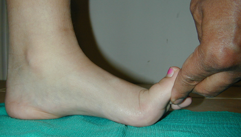
As chiropodists/podiatrists, we perform structural and biomechanical evaluations every single day. We watch patients walk, evaluate force distribution, palpate painful insertions, and routinely reach down to perform a passive toe-raising maneuver.
In clinical shorthand, we call this Jack’s Test, or the Hubscher maneuver. We frequently treat it as a quick, binary confirmation of whether a patient’s windlass mechanism is structurally intact.
However, looking back at the original mid-twentieth-century literature, and pairing it with innovative contemporary neuro-biomechanical research, reveals that this test was never meant to be a simple yes-or-no screening tool. Critical modern insights reveal how your manual exams can accurately pinpoint exactly where an arch is failing across all three anatomical planes.
The Engineering Basis: Hicks’ Windlass
To understand Jack's work, we must pair it with the foundational biomechanical research conducted around the same time by J.H. Hicks. In his classic 1954 study, Hicks modeled the human foot as a triangular architectural truss.
Within this framework, the tarsal and metatarsal bones comprise the rigid compression struts, while the plantar aponeurosis operates as the tensile tie-rod, or bowstring, spanning the base of the medial longitudinal arch.
When a patient moves into terminal stance and pre-swing, extension of the metatarsophalangeal joints (specifically the hallux) winds the plantar fascia around the mechanical drum of the metatarsal heads. This winding action shortens the effective length of the aponeurosis, automatically hoisting the calcaneus anteriorly, inverting the rearfoot, externally rotating the tibia, and packing the midtarsal joints into a rigid lever optimized for propulsion.
Dr. Jack’s True Intention: Mapping out the Flatfoot Deformity
A year before Hicks published his definitive windlass model, orthopedic surgeon Ewen A. Jack was grappling with a massive clinical problem in pediatric orthopedics: how to properly classify and treat pediatric flatfoot.
Writing in the Journal of Bone and Joint Surgery in 1953, Jack sought a definitive, non-invasive method to distinguish between a flexible, postural flatfoot and a structurally rigid, fixed deformity.
He did not just want to see if an arch would appear. Jack was designing a precise pre-surgical diagnostic stress test intended to do two things:
1. Differentiate Flexible from Rigid Pathology
Jack realized that manually dorsiflexing the hallux in a weight-bearing position used the foot’s anatomy to challenge the joints. If the arch successfully restored itself and the heel inverted under this passive leverage, it proved to the surgeon that the joints of the midfoot retained the dynamic capacity to correct their alignment. If the arch remained completely collapsed under maximum manual force, it flagged a rigid, structural pathology, such as a tarsal coalition, that standard conservative therapies could not fix.
2. Identify the Exact Structural Break under Radiograph
Jack’s true brilliance lay in combining the toe-raising test with weight-bearing radiographs. By comparing standard standing X-rays with X-rays taken while manually lifting the big toe, he categorized flatfoot deformities based on the specific joint where the structural column buckled:
The Naviculo-Cuneiform Break: If the toe-raising test cleanly restored the arch profile on the table and aligned the bones under X-ray, Jack interpreted this as localized joint instability specifically at the naviculo-cuneiform interface, while the proximal talonavicular joint remained stable.
The Perpendicular Talus Failure: If the test failed to lift the midfoot, Jack noted that a severely plantarflexed or vertical talus created a mechanical block. The foot simply lacked the necessary structural leverage to force the navicular back under the dropped head of the talus.
For patients who demonstrated a clear, correctable naviculo-cuneiform break, Jack used the positive result of his toe-raising test as the primary indicator that the patient was an ideal candidate for a naviculo-cuneiform fusion to permanently stabilize that specific column.
Expanding the Clinical Triad: Isolating the True Failure Point
To gain a flawless diagnostic picture of a collapsing foot, we must look past the sagittal plane of Jack's test and build a complete clinical triad. By pairing Jack's test with the single-leg heel raise and Pasapula's neutral heel lateral pull/push test, we can isolate whether a failure is skeletal, neuromuscular, or ligamentous.
| Diagnostic Test | Primary Mechanism Evaluated | Primary Tissue Questioned | Clinical Value |
| Jack's Test | Passive Windlass (Sagittal Plane) | Plantar fascia integrity and midfoot joint mobility | Determines if the skeletal and fascial architecture can physically lock without muscular assistance. |
| Single-Leg Heel Raise | Active Concentric Pull (Frontal Plane) | Tibialis posterior tendon and gastroc-soleus drive. | Evaluates if the tendon can actively invert the heel and lift the arch against gravity during propulsion. |
| Pasapula Neutral Heel Lateral Push Test | Joint Laxity (Transverse Plane) | Superomedial calcaneo-navicular (Spring) ligament complex. | Isolates whether a hidden spring ligament tear is causing the talar head to escape medially, ruining the structural truss. |
By utilizing this triad, your exam becomes incredibly precise. For example, if a patient presents with a positive Jack's test but a failed single-leg heel raise, you have isolated a active motor problem, pointing heavily toward tibialis posterior tendon dysfunction.
However, if Jack's test fails and you elicit prominent hypermobility or pain during Pasapula's neutral heel lateral pull test, the windlass isn't working because the underlying structural floor has given way. The spring ligament is lax, allowing the talar head to displace medially, which completely de-tensions the plantar fascia winch.
The Modern Paradigm: Moving from Passive Strings to Active Motors
While Hicks and Jack gave us a brilliant geometric foundation, contemporary research from the labs of Dr. Luke Kelly and Dr. Gabriel Moisan proves the foot is far from a passive, mechanical winch.
The Active Buttress (Luke Kelly Group)
Luke Kelly’s team proved that passive strain on the plantar aponeurosis via toe extension cannot fully explain the rapid foot stiffening observed during gait. Using in vivo loading and electromyographic data, they demonstrated that foot stiffening during push-off is actively modulated by contractions of the ankle plantarflexors and the plantar intrinsic foot muscles, including the abductor hallucis, flexor digitorum brevis, and quadratus plantae. The arch is an active, dynamic buttressing system. A failed Jack's test on your clinical table might highlight a neurological or muscular deficit in the patient’s intrinsic capacity to stabilize their own arch, rather than a simple mechanical jam.
Individualized Kinetics (Gabriel Moisan Group)
Gabriel Moisan’s research warns us against oversimplifying pathologies like plantar fasciopathy as basic, uniform windlass failures. His quantification of first MTPJ dorsiflexion and supination resistance shows massive individual variation. In conditions like Progressive Collapsing Foot Deformity, orthotic success is not about fixing a broken passive string. Instead, orthoses work by altering kinetic resistance to assist active muscular function, reducing the physical workload required by the tibialis posterior and intrinsic muscles.
The Clinical Breakdown: What the Test Tells Us on the Table
While modern podiatric practitioners rarely use Jack’s test to plan an arthrodesis, the mechanical forces highlighted by both historical and modern literature are the exact same ones causing our patients' musculoskeletal complaints today.
When you perform the maneuver in your clinic, you are assessing two distinct functional phases:
1. Functional Hallux Limitus (The Jammed Lever)
If a patient has adequate passive hallux dorsiflexion non-weight-bearing, but Jack’s test fails to elicit arch restoration weight-bearing, you are witnessing an immediate failure of the windlass mechanism. As established by Dananberg, this sagittal plane restriction leaves the midfoot an unstable, flexible structure during propulsion, forcing the intrinsic musculature and the tibialis posterior tendon to work past their physiologic capacity to stabilize the medial column.
2. Plantar Fasciopathy (The Overloaded Tie-Rod)
Ewen Jack himself noted a critical caveat that every modern clinician should remember: the efficiency of the toe-raising test is easily overpowered by a tight Achilles tendon.
When an Achilles equinus is present, the severe posterior pulling force overpowers the passive mechanical advantage of the hallux windlass. This creates a violent tug-of-war across the plantar aspect of the foot, focusing massive tensile strain directly at the medial calcaneal tubercle. This is the exact mechanical etiology behind chronic, recalcitrant plantar fasciitis.
Prescription Logic: Orthotic Modifications for a Failed Jack's Test
When Jack’s test is delayed or completely absent on the clinical table, standard, geometric arch support is rarely enough. The orthotic prescription must be engineered to actively lower the mechanical barriers blocking the windlass mechanism or to artificially provide the stabilization the plantar fascia cannot generate.
Consider deploying these targeted modifications based on your palpatory and visual findings:
First-Ray Cutout and Kinetic Wedge/Reverse Morton’s: If the first metatarsal head is structurally elevated or jammed, ground reaction forces will block the hallux from dorsiflexing. A localized cutout under the first MTPJ, often paired with a high-density EVA kinetic wedge under the remaining four metatarsal heads, allows the first metatarsal to plantarflex natively, dropping the drum of the winch and initiating an immediate, timely windlass engagement.
Kirby Medial Heel Skive: When an absent Jack's test is accompanied by a severe, high-velocity rearfoot eversion, the subtalar joint axis is pushed heavily medially. An aggressive medial heel skive increases the supinatory torque medial to the subtalar joint axis. This physically reduces the pronatory workload on the medial column, making it easier for a weak windlass mechanism to operate.
Pasapula-Driven Midfoot Stabilization: If your exam revealed a transverse plane failure via a positive Pasapula test, your orthotic must prioritize stopping the medial migration of the talonavicular joint. Incorporating a deep heel cup, a high medial flange, and a rigid shell material prevents the structural collapse of the spring ligament, holding the struts in place so the windlass mechanism actually has a stable foundation to wind against.
Summary of Clinical Takeaways
To maximize the diagnostic value of Jack's test in your daily practice, keep these five central pillars in mind:
Original Intent: Designed by Ewen Jack in 1953 as a pre-surgical radiographic stress test to differentiate flexible from rigid flatfoot and pinpoint the exact naviculo-cuneiform break for joint fusion.
The Diagnostic Triad: Pair Jack's Test (passive sagittal stability) with the Single-Leg Heel Raise (active frontal drive) and Pasapula's Neutral Heel Lateral Pull Test (passive transverse/spring ligament integrity) to immediately identify the true root of a collapsing foot.
The Mechanical Synergy: Bending the toe activates Hicks' 1954 passive windlass mechanism, but modern science from the Kelly and Moisan groups proves this works in strict synergy with an active, neuromuscular buttressing system driven by the foot's intrinsic musculature.
The Achilles Roadblock: A positive or negative Jack's test is heavily dictated by the presence of an Achilles equinus. Gastroc-soleus tightness will violently overpower the mechanical leverage of the big toe.
Targeted Prescriptions: Use your multi-planar findings to dictate specialized orthotic modifications, utilizing first-ray cutouts to unlock sagittal plane motion, medial heel skives for frontal plane eversion, and deep heel cups with high flanges to support a failing spring ligament.
In a future blog I will outline the research work of the Pasapula research group.
My next blog post I will discuss the foot posture index.
References
1. Jack EA. Naviculo-cuneiform fusion in the treatment of flat foot. J Bone Joint Surg Br. 1953 Mar;35-B(1):75-82.
2. Hicks JH. The mechanics of the foot: the plantar aponeurosis and the arch. J Anat. 1954 Jan;88(1):25-30.
3. Dananberg HJ. Gait style as an etiology to chronic postural pain. Part I. Functional hallux limitus. J Am Podiatry Med Assoc. 1993 Aug;83(8):433-41.
4. Kirby KA. The medial heel skive technique: improving pronation control in foot orthoses. J Am Podiatry Med Assoc. 1992 Apr;82(4):177-88.
5. Kelly LA, Cresswell AG, Farris DJ. Foot Stiffening during the push-off phase of human walking is linked to active muscle contraction, and not the windlass mechanism. J R Soc Interface. 2020 Jul;17(168):20200258.
6. Kelly LA, Lichtwark G, Farris DJ, Cresswell A. Shoes alter the intrinsic mechanics of the foot’s longitudinal arch during walking. J R Soc Interface. 2020 Jan;17(162):20190804.
7. Moisan G, Chicoine D, McBride S, Griffiths IB. Supination resistance variations in foot and ankle musculoskeletal disorders: implications for diagnosis and customised interventions with wedged insoles. J Foot Ankle Res. 2023 Dec;16(1):82.
8. Robb K, Ryan M, Moisan G. Clinical Outcomes of Custom Foot Orthoses in Progressive Collapsing Foot Deformity: A Retrospective Cohort Analysis. J Clin Med. 2026 Apr;15(8):2104.
9. Pasapula C, Devany J, Chalasani P, et al. The neutral heel lateral push test: a clinical examination tool for spring ligament laxity. Foot Ankle Surg. 2024 Feb;30(2):142-148.